| GISdevelopment.net ---> AARS ---> ACRS 1991 ---> Poster Session |
Steady-State Leser
Fluorometry in Sea-Water Phyto – and Zooplankton analysis
Andrey A. Demidov Elina A.
Chernyavskaya
Moscow State University, Physics Department
Moscow 119899, Lenin Hills, USSR
Moscow State University, Physics Department
Moscow 119899, Lenin Hills, USSR
Abstract
There is developed the new promising methods for the plankton investigations based on the steady-state He-Cd laser ( l=440 nm). This methods uses the fluorescence of phyto – and zooplankton pigments excited by a continuous laser light. There is detected simultaneously the water Raman signal as a calibration signal. Developed methods was tested in a laboratory as well as in a native sea-water conditions ( South Atlantic). They can be used in analyzing of intact algae in detect chlorophyll a concentration in native intact algae up to 10 nanogram/liter without concentration a probe; b) to estimate the DCMU; c) to detect chlorophyll a and pheophytin a concentrations in acetone extracts of phyto – and zooplankton up to 1 nonogram / liter; d ) to estimate the photosynthetic activity of algae without using of any chemicals like DCMU; c) to detect chlorophyll a and pheophytin a concentrations in acetone extracts of phyto – and zooplankton up to 1 nanogram / liter; d) to control the feed process of individual sample of zooplankton. Created He-Cd fluorosensor has very simple construction. It is cheap and easy in use.
Introduction
Laser remote sensing method are widely use now in the ocean logy practice, particularly, in the phytoplankton chlorophyll analysis [ 1-6]. Commonly, they use the powerful laser pulses to excite the algae pigments fluorescence which carries on the information of a phytoplankton. The most popular lasers in these investigations are YAG: Nd+3 laser ( 8 = 532 nm), nitrogen laser ( 337 nm) and dye lasers (tunable wavelength ). the use of lasers enables one to develop express, distant, non contact and nondestructive methods for a phytoplankton investigations.
In our research we try to use the steady-state He-Cd laser (l=440 nm). Our methods are based on the principles and ideas developed before for the YAG laser application for the phytoplankton and zooplankton pigments measurements [ 3-7]. The use of YAG laser fluorosensor enables one to measure the intact algae chlorophyll a (chl a ) fluorescence in native water ( without any concentration procedure ) with a high biological productivity down to the mesotrophic waters, but it doesn’t work in the oligotrophic waters because of a not sufficient sensitivity.
We hoped that this problem can be overcomed with the use of the He-Cd laser because this laser can more efficiently excite the chl a fluorescence ( in vivo ) as well as in vitro ) than in the case of YAG laser. The He-Cd laser light is absorbed by chl a in the Soret line, Which has the maximum absorption coefficient. Besides, in this case the Raman signal (IR) and chl a fluorescence (If1) are better separated and as a result the smaller ratio F=If1/IR can be achieved ( see below ). All these facts increase the sensitivity of our laser fluorometry method {8}.
Materials and Methods
In Our research we have studied a lot of different sea-water plankton objects:
- About 60 probes of a different algae cultures ( Gymnodinium, Sceletonema costatun, Prorocentrum micans, Platymonas viridis ) – laboratory experiments;
- Acetone extracts of marine phytoplankton ( from the same bathometers ) that was prepared and controlled in accordance with standard method [ 9-11] – in vitro experiments;
- acetone extracts of marine phytoplankton ( from the same bathometers ) that was prepared and controlled in accordance with a standard method [9-11] – in vitro experiments;
- hundreds of acetone extracts of a countable number ( up to one sample ) of zooplankton samples that was made in accordance with the work [7].
In this figures the ‘short wavelength ’ signal (IR) is the Raman scattering of a laser light by water ( Fig. 2) or acetone ( Fig.3) molecules; the ‘long wavelength’ signal (If1) is the pigments fluorescence ( for example, ch1 a, fluorescence). In the words [4-6] there were proofed that the ‘fluorescence parameter’ F=If1/IR is independent on the experiment conditions ( the internal calibration principle, IR calibration signal ) and is linearly connected with the fluorescent pigments concentration C = "M, where " is a parameter. This linear dependence takes place if the because of that the algae functional state can’t influence to the " parameter.
Phytoplankton Photosynthetic Activity Diagnostic
Laser light has enough intensity to influence to the process of a closure of algae photosynthetic reaction centers (RC). and it can be used in the algae photosynthetic activity estimation without involving of any chemicals like DCMU. As we have shown in our research, algae fluorescence behaviors is connected with the laser photo induced driving of a functional states of the algae photosynthetic RC. In turn the RC opf algae ch1 a fluorescence. The physical mechanism of this non-linear phenomena is very simple. When r<10-8 ( Fig.1, ‘ low intensity’ region ) the rate of a laser photons absorption by a ch1 a molecules of an algae light – harvesting antenna is less then the rate. Of Exactions “Utilization” In The RC and then F=Fmin. Here, r=4F, where 4 is the free ch1 a molecules excitation decay time, F is the laser light absorption cross-section, F is the laser light beam photon density, [ photon. sm. s. }. As soon as this balance turns over RC
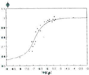
Would be locked by excitons utilization procedure and reaction centers couldn’t efficiently trap antenna excitions and fluorescence would rise and achieve M=M at the r > 10-6 ( ‘high intensity’ region).
In the ‘high intensity’ mode the value of a fluorescence parameter (Fmax) isn’t influenced by a initial algae functional state but in the ‘low intensity’ mode this dependence takes place. So the ration Fmax /Fmax will characterize the algae photosynthetic activity.
Determination of A sea-water chlorophyll a concentraction in the intact phytoplankton
When intact phytoplankton is illuminated by a He-Cd laser its fluorescence is the fluorescence of a ch1 a molecules contained in this algae. It may be proofed that a ch1 a concentration C ch1a = "int. F, where "lint. at the 8=440 nm is equal
Here
 is the ratio of ch1 a and water molecular
weights;
is the ratio of ch1 a and water molecular
weights;  is the Raman cross-section
(Stock’s shift v=3440 sm-1);
is the Raman cross-section
(Stock’s shift v=3440 sm-1);  is the concentration of a water molecules;
D8f1/D8R is the ratio of half widths of the fluorescence and Raman
signals;
is the concentration of a water molecules;
D8f1/D8R is the ratio of half widths of the fluorescence and Raman
signals;  is the effective fluorescence
cross-section at the 8=440 nm [8]. Then we obtain:
is the effective fluorescence
cross-section at the 8=440 nm [8]. Then we obtain: 
During the ‘VITYAZ’ trip we have analyzed a set of intact sea-water phytoplankton probes (about 30) to find exp. The typical spectrum aint presented in the Fig. 2. Ch1 a concentrations was measured by a standard spectrophotometric method [9] and M - by our fluorometric method. We have got the linear dependence between Cchla and M.
With the correlation coefficient R (CchlaF = 0.93). This result agrees well with our theoretical prediction (2).
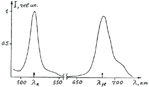
Fig.2. Response signal of a natural intact sea-water phytoplankton; l – laser light Raman scattering by a water molecules, lf1 – phytoplankton ch1 a fluorescence. The same result was obtained in a laboratory investigations of algae cultures ( Gymnodinium, Sceletonema costatun, Prorocentrum micans, Platymonas viridis ) in various moments of their growing. We studied about 60 probes using the OMA-1 based laser fluorometer. For these algae we have got exp. The typical spectrum "
 consequently for the Gymnodinium,
Sceletonema costatun, Prorocentrum micans, Platymonas viridis.
consequently for the Gymnodinium,
Sceletonema costatun, Prorocentrum micans, Platymonas viridis. Our method has allowed us to measure the ch1 a concentration in an intact algae up to 10 nanogram/literand that ‘opens the door’ to the phytoplankton laser fluorometry in the oligotrophic areas of oceans.
Determination of The Chlorophyll a and Pheophytin a Concentrations In The Phyto-and Zooplankton Acetone Extracts
One the base of principles described above we have developed the extract method of detection of extremely low concentrations of ch1 a and pheophytin a (ph a) in the acetone extracts. In this method we used the well known acidification technique [9]: the extract fluorescence (Fig.3) was measured twice – before (M) and after (Ma) its acidification by a HCl acid [5,6]. In this case we took the acetone Raman signal (v=2970 sm-1 8R=506 nm, see Fig.3) as the calibration signal (IR).
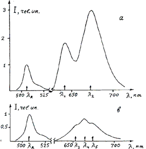
Fig. 3. Response signals of a phytoplankton acetone extract before (a) and after (b) its acidification by a HC1; 8R- laser light Raman scattering by a acetone molecules, 8285 – fluorescence of ch1 a and ph a, 8183, 84 – fluorescence of chlorophylls b, c and their pheophytins.
The extract response signal (Fig. 3) are complex both before and after acidification. Here there are not only the ch1 a and ph a fluorescence but also the fluorescence of chlorophylls b, c (ch1 b,c ) and their pheophytins (after acidification only pheophytins ). In the case of a zooplankton extracts, sometimes, there were fluorescence of some unrecognized ‘gut’ pigments. On the one hand it makes more
Difficult to select ch1 a and ph a signals and decreases the accuracy of their concentrations estimation but on the other hand fluorescence spectra contain a lot of information. This information can be used in the simultaneous ch1 a,b,c, and ph a,b,c, and ph a,b,c, estimation and we plan to develop the modified method to do that. Following the works [5,6] we have got the theoretical expression for the C chla and Cpha in the 90% acetone-water solution:
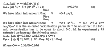
This extract method has the ch1 a determination sensitivity up to 1 nanogram/liter. That was allowed us to study pigments in the extremely low concentrated probes and in the guts of individual zooplankton samples. If one would prepare the 1 ml of extract from the one sample of a zooplankton it will be possible to estimate the pigments contain in its gut as low as 12-12 gram. So you can investigate the feed process of an individual zooplankton sample.
References
- Measure R.M. ( 1984 ) Laser Remote Sensing . Ed. John wiley & Sons.
- Hoge F.E. and R.M. Swift ( 1989) Remote sens. Environ,. V.30, pp.67-76.
- Fedeev V.V., A.A. Demidov and A.M. Chekalyuk (1991) This conference.
- Demidov A.A., A.M. Chekaluk, T.V. Lapshenkova and V.V. Fadeev ( 1988) Meteorologiya I Gydrologiya (USSR), N 6, pp. 62-70.
- Demidov A.A. E.V. Baulin, V.V,. Fadeev and L.A. Shur ( 1981) Okeanologiya (USSR), v-21, pp.174-179.
- Demidov A.A. and I.G. Ivanov ( 1989) Izvestiya akademii Nauk SSSR, seriya biologycheskaya (USSR), v3, pp. 378-856.
- Pasternak A.F., A.A. Demidov, A.V. Dritz and V.V. Fadeev ( 1987)
- Demidov A.A.and E.A. Chernyavskaya ( 1991) Biofozika (USSR) in press.
- Water. Spectrophotometric method of a chlorophyll a determination. – GOST USSR 17.1.04.02-90, Moscow, - 14 p..
- UNESCO ( 1966) SCOR-UNESCO working group 17, pp. 9-18.
- Jeffrey S.W. and G.F. Humphry ( 1975) Biochem. And Physiol Planz. Bd. 167, pp. 191-194.
- Valkunas L., N.E. Geacintov, L.France and J. Breton ( 1991) Biophys. J., v-59, pp. 397-408.