| GISdevelopment.net ---> AARS ---> ACRS 1991 ---> Oceanography |
Laser Remote Sensing of
Sea-Water Plankton
Victor V. Fadeev, Andrey A.
Demidov,Alexander M. Checkalyuk
Moscow State University, Physics Department
Moscow 119899, Lenin Hills, USSR
Moscow State University, Physics Department
Moscow 119899, Lenin Hills, USSR
Abstract
Our group have been working the problem of laser remote sensing of sea-water phytoplankton since 1975. In this way we have investigated the fundamental problems of a laser light propagation in natural waters and its interaction with the intact algae. Now we have a great experience in this field in theoretical as well as in practical approaches n about 20 shipboard expeditions in Pacific, Atlantic, India oceans and in a lot of seas. Our methods are based on an analyzing of chlorophyll fluorescence of intact phytoplankton and Raman signal of water molecules which is used as internal calibration parameter. Both signal are detected by lidar (laser fluorosensor) consisted of impulse YAG: Nd-laser (l=532 nm) and Optical Multichannel Analyzer (OMA). Lidar work in the remote mode as well as in the probe testing mode. This lidar was successfully used in quantitative search of concentrations of a phytoplankton chlorophyll in different areas of World Ocean down to mesotrophic water (medium biological productivity) and we got a log of fine structure maps (space resolution on a broad areas. Now we develop some new promising methods of detecting of the photosynthetic activity and shown the rate of its influence on the lidar detecting of phytoplankton fluorescence. High sensitivity of our fluorometric methods (up to 0.1 mg/1 of chlorophyll detection in natural waters and up to 0.1 mg/1 in acetone extracts of plankton) enabled us to develop the new method of zooplankton feed process analysis up to the level of individual sample.
Introduction
In the ocean investigations, one of the prameters broadly used in practice is the phytoplankton chlorophyll a (chl a) fluorescence. This fluorescence can be used as a quantitative parameter for the estimation of a sea-water biological productivity. Recently in the ocean logy practice, laser fluorometers have been involved [1-16]. They are mainly used in the laser remote sensing of a sea-water. This can be explained by the specific features of the laser sources that can produce a very powerful monochromatic beam with a low divergence. Among the groups working in this area is the Group of Water Laser Remote Sensing of the Physics Department of Moscow State University [1-7]. The researches of this group have developed some sea-water Remote Sensing methods as well as investigated the laser induced fluorescence of intact algae in the sea-water probes [1-5], fluorescence of phytoplankton pigments in extracts [2,7] and fluorescence of pigments in the zooplankton extracts [6].
The specific properties of the laser light open up some new fundamental potentialities of fluorometry that are hardly possible in the “classic” lamp fluorometry. One of these properties is the fixed light wavelength that provides for a stability of absorbance and fluorescence in the case of using different laser fluorometers but with lasers of the same type. It also enables an easy detection of the Raman scattering of the laser light by the solvent molecules that can be used as an internal standard (1, 3, 10, 14, 15).
Regarded as the pioneers in suing the Raman signal as an internal standard in sea-water analysis are D.A. Leonard and C.H. Chang (1974) (14), M. Brown (1974) (15) and V.V. Fadeev (1975) (1). In their works they investigated the fluorescence of intact phytoplankton with using water Raman signal. Then in 1978 this method was surely proved mathematically by Klyshko and Fadeev (3). Since 1974 it has been widely used in the laser remote sensing of natural sea-water.
Laser Fluorometer (LIDAR) Construction
In our investigations, we have used the shipboard mounted lidar system developed n the Moscow University which can be easily accommodated to the Remote Sensing mode or to the probe analysis (1-7). Used as the laser source is the impulse YAG laser (l=532 nm). t~ 10 ns, power p~500 KWt. Lidar was positioned in the ship laboratory. Distance from the lidar to the sea surface was about 10-15 m. The laser beam illuminates the sea surface after reflection from the mirror mounted on the ship false board or the cuvette with the water probe of phytoplankton to excite the phytoplankton fluorescence (lf1@685 nm ) and water Raman scattering (lR=651 nm). Both signals are detected by an Optical Multichannel Analyzer (OMA, PARC production, USA). The OMA detector is gated by high voltage pulses of 0.04-1 ms duration synchronized with the laser light pulses. This approach enables one to do measurements under natural conditions of the sunshine and it would not influence the results of measurement.
The whole spectrum is stored in the OMA memory to be processed by a computer interfaced with it. Besides, this computer presets the mode of operation of the laser fluorometer as a whole. OMA operates in the mode of parallel detection of the spectrum (l=275 nm) and it is effective in detection of fluorescence intensity that is 1012 - 1013 times less than the laser light intensity. In fact, in the remote sensing mode we measure the average value of fluorescence of the upper 2-5 m of water because of a great absorbance of response signal (l=650-690 nm) by a natural sea-water.
The use of a computer enabled us to do advanced automatization of a whole lidar complex and process of measurements. As a result, we have achieved the time duration of a one measurement circle (from the start to the final result) about 1.5 min. That allowed us to obtain the space resolution of a sea-water monitoring about 450 m at a ship speed 10 miles/hours. This is not a principle limit and we know how to improve this result.
Laser Fluorometry of Phytoplankton and its Applications
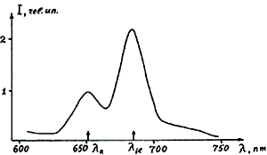
In the Fig. 1 there is presented the typical spectrum of detected response signal. There, one can see two signals: (1) Raman signal (IR lR=651 nm) and (2) phytoplankton chl 1 a (If1, lf1 @685 nm). Both signals are similarly influenced by the measurement conditions (the form of water surface or the cuvette positions, illumination conditions etc.) and their ratio f=If1/IR will not be dependent on these conditions and, consequently, on the fluorometer type (3). We name f as ‘fluorescence parameter’. This parameter is a quantitative value of pigment fluorescence and it is linearly connected with the pigment concentration f=aC (2, 3, 5), where C is the concentration and a - coefficient. In fact, Raman signal is regarded as an internal standard to be used for the fluorescence calibration.

Fig. 1. Typical spectrum of response signal detected in South Atlantic near Namibia, 1985-1986, the 43rd scientific trip of the ‘Academitian Kurchsatov’ ship (Institute of Ocean logy, USSR Academy of Sciences); lR – water Raman, l f1 – phytoplankton chl a fluorescence.
One of the “underwater reef” in the laser fluorometry of a phytoplankton is the effect of fluorescence saturation when any one use the powerful laser light to excite this fluorescence (2, 5). This effect is connected with a set of specific processes in the pigment light-harvesting antenna of algae, for example, with the singlet-singlet annihilation in it. So, one have to do special precautions to avoid it or to do a necessary correction procedure (2, 5).
In the sea-water monitoring there is necessary to know not only the intensity of a phytoplankton fluorescence but mainly the value of a chl a concentration C (C=af). We have been investigating the problem of a estimation for years, now it isn’t yet solved for sure. But in a set of scientific expeditions in the Pacific, Atlantic and Indian oceans and in some of seas we have got:

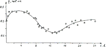
Where f0 is the fluorescence parameter f intact phytoplankton fluorescence measured in the probes in the shipboard laboratory with the specific laser excitation mode and with the correction procedure of fluorescence saturation elimination (2, 4, 5). This method allows one to measure chl a concentration as low as 0.1 mg/1. The other one “underwater reef” in the laser Remote Sensing of a natural phytoplankton is the day time dependence of a phytoplankton fluorescence connected with a natural day tie changing of an angle photosynthetic activity. This effect causes the day time modulation of the fluorescence responses single (see Fig. 2) and if any one wants to do correct monitoring of a sea-water subsurface phytoplankton fluorescence he has to d the appropriate correction procedure. We have to mention that this day time dependence (the curve form and value of the modulation coefficient is the function of a phytoplankton environment inhabitance and average sunshine illumination intensity. Now we have some laser methods of estimation of the algae photosynthetic activity (see materials of this conference).

Fig. 2. Day time dependence of a normalized fluorescence of subsurface phytoplankton (in fact, fluorescence quantum yield of intact algae in situ); remote sensing on station near the Elephant island, South Atlantic, 1985-1986.
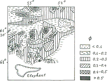
A special interest of using the laser monitoring in the sea-water control is connected with its possibility to get express-information of a phytoplankton chl a distribution on the large areas in a distant mode. Our lidar enables on to do scanning of a sea surface with a high resolution (up to hundred meters) at a ship speed over 10 miles/hour and analyze the fine structure of a phytoplankton fluorescence distribution. For example, we have found that variability of phytoplankton fluorescence can achieve about 300% on the 1 mile duration. Now we have got a set of maps of phytoplankton fluorescence distributions over the large areas of some oceans and seas. One of them is presented in the Fig. 3.

Fig. 3. Subsurface phytoplankton fluorescence distribution map, near the Elephant island, South Atlantic, 1985-1986.
This map (area about 60x60 miles) was obtained during a week involving the correction to the fluorescence day time dependence measured on the one station near the Elephant island (see Fig. 2). When the ship came to this field after 9 days the fluorescence distribution pattern was the same. Moreover, in the points of crosses of old and new courses the fluorescence intensities were the same with the accuracy better than 20% (day time correction procedure was involved in the both cases).
Laser fluorometry of acetone extracts of chlorophyll a and pheophytin a of a phyto-and zooplankton
In some cases there is necessary to analyze not intact algae but their extracts, for example, in the case of extremely low pigment concentration and limited volume of an initial water probe (2) or in the case of zooplankton extract (6). If any one prepare the acetone-water extract (90% acetone) of sea-water phytoplankton with accordance with the standard procedure [17-19] or zooplankton extract, it is possible to measure partial concentration of chlorophyll a and pheophytin a (py a) in it:

Here we use the same idea of calibration by a Raman Signal, but now we use the acetone Raman (l=630 nm); f0 and fa0 are the fluorescence parameters before and after extract acidification by a HCI acid (it is well know acidification technique [17-19]); achla = 3 mg/1, apha 1.5 mg/ (our last estimation); ‘acidification parameter’ z = achl a / aph a = 2. The accuracy of these formulas is about 20%. It enables one to measure the chl a and ph a concentrations up to 10 nanogram/liter.
The high sensitivity of this method makes a reality the efficient investigation of a zooplankton feed process and analyzing of extremely low concentrated probes.
References
- Fadeev V.V., L.B. Rubin, S.V. Kust, V.S. Petrosyan, V.G. Tunkin and L.A. Haritonov (1980) – In: ‘ Complex investigations of a ocean nature’ – Report in the 1st Conference of Moscow State University on World Ocean Problems, Moscow, December 1975. Moscow (USSR), pp. 213-217.
- Demidov A.A., E.V. Baulin, V.V. Fadeev and L.A. Shur (1981) Okeanologiya (USSR), v. 21, pp. 174-179.
- Klyshko D.N. and V.V. Fadeev (1978) Dokl. Akad. Nauk SSSR, v. 238, pp. 320-323.
- Demidov A.A., A.M. Chekalyuk, T.V. Lapshenkova and V.V. Fadeev (1988) Meteorologiya i Gydrologiya (USSR), N 6, pp. 62-70.
- Fadeev V.V., A.A., Demidov, D.N. Klyshko, O.I. Koblentz-Mishke and V.M. Fortus (1980) Trudy Instituta Okeanologii Akademii Nauk SSSR, v. 90, pp. 219-235.
- Pasternak A.F., A.A. Demidov, A.V. Dritz and V.V. Fadeev (1987) Okeanologiya (USSR), v. 27, pp. 852-856.
- Demidov A.A. and I.G. Ivanov (1989) Izvestiya Akademii Nauk SSSR, seriya biologycheskaya (USSR), v. 3, pp. 378-385.
- Bunkin A.F. and A.L. Surovegin (1986) Okeanologiya (USSR), v. 26, pp. 532-536.
- Arverson J.C., J.P. Millard and E.C. Weaver (1973) Astronautia Acta, v. 18, pp. 229-239.
- Hone F.E. and R.M. Swift (1981) Appl. Opt., v. 20, pp. 3197-3205.
- Hone F.E. and R.M. Swift (1989) Remote Sens. Environ., v. 30, pp. 67-76.
- Kim H.H. (1973) Appl. Opt., v. 12, pp. 1454-1459.
- Mumola P.B., O. Jr. Jarrett and C.A. Jr. Brown (1973) In: Second Joint conference on sensing of environmental pollutants. Washington., D.C., pp. 53-63.
- Leonard D.A. and C.H. Chang (1974) United State Patent: 3,806, 727. Apr. 23.
- Brown M. (1974) Report N 29, Kobenhavns universtitet Institute for fysisk oceanografi, Copenhagen, December 1974. – 29 p.
- EARSEL Conference ‘Lidar Remote Sensing of Land and Sea’ (1991) Florence (Italy), May 6-8.
- Water. Spectrophotometric method of a chlorophyll a determination. – GOST USSR 17.1.04.02-90, Moscow, - 14 p..
- Determination of phtosynthetic pigments in sea water . UNESCO (1966) SCOR-UNESCO Working group 17, pp. 9-18.
- Lorenzen C.J. (1967) Limnol. Oceanogr., v. 12, pp. 343-346.